
Patient Overview
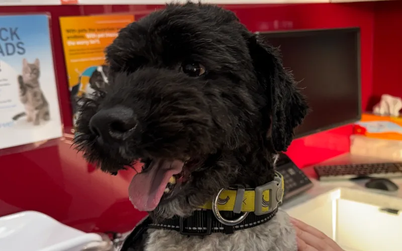
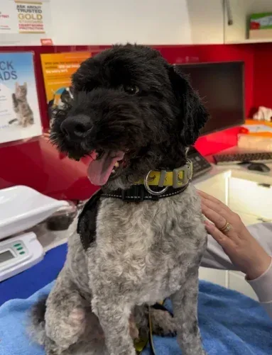
When Barney, a six-year-old miniature poodle cross, arrived at Casey and Cranbourne Veterinary Hospital, his family knew something was not right.
Over four days, he had become increasingly lethargic, had stopped eating, and had vomited twice. Despite normally enjoying a diet of cooked chicken, rice, vegetables and dry food, he had shown virtually no interest in food since Sunday evening. He was also drinking very little water and seemed uncomfortable.
Barney’s family were understandably concerned. He was a much-loved member of the family, carrying special significance after the loss of their father, and they knew his behaviour was not normal.
“If your pet just isn’t quite right, trust your instincts. Subtle changes can sometimes be the first sign of something more significant.”
Looking Beyond The Obvious
When Dr Elizabeth Doran examined Barney, several possible causes needed to be considered. His symptoms could have been related to pancreatitis, Addison’s disease, gastrointestinal upset, a foreign body obstruction, or other underlying medical conditions. While his examination was largely unremarkable, his abdomen was tense and uncomfortable when palpated, suggesting an abdominal source of pain.
Given the broad range of possible causes, a thorough diagnostic work-up was recommended. We started with comprehensive in-house blood testing, including a canine-specific lipase test used to help assess for pancreatitis.
The results confirmed elevated pancreatic enzymes consistent with pancreatitis. Importantly, his kidney and liver values, electrolytes, and red and white blood cell counts were largely within expected ranges, helping us rule out several other conditions.
At this stage, Barney’s diagnosis appeared relatively straightforward. However, his clinical progress over the following 24 hours suggested there was more to the story.
A Change In The Clinical Picture
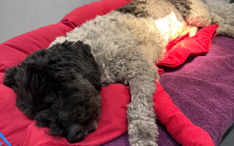
Despite supportive care, Barney remained painful and continued refusing food. He was admitted to the hospital for ongoing monitoring, intravenous fluids and pain relief. While being cared for by our veterinary nursing team, a significant change was noticed. Barney’s urine had become unusually dark brown.
Further testing revealed bilirubin and blood in the urine, while repeat blood work showed mild anaemia and evolving changes warranting further investigation. Although pancreatitis had been confirmed, these new findings suggested another process may also be occurring.
This is where ongoing reassessment becomes important. In complex cases, diagnosis is not always a single event. New information can emerge as a patient’s condition evolves.
Advanced Imaging Reveals Two Significant Findings
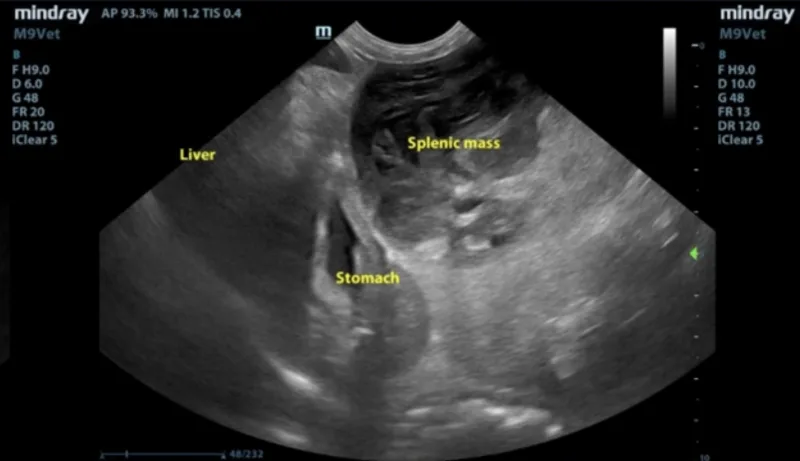
To investigate further, an abdominal ultrasound was arranged with visiting veterinary ultrasonographer Dr Lisa Muscat from In Focus Imaging.
The ultrasound revealed an unexpected finding. Barney had a large eight-centimetre mass on his spleen, along with a small amount of free fluid within the abdomen consistent with minor bleeding from the splenic lesion.
A second, even more unusual, finding was also identified. A large abnormal blood vessel connection was seen involving Barney’s caudal vena cava, one of the body’s major veins. While the ultrasound clearly demonstrated the vascular anomaly, it was not possible to determine exactly where the vessel was connecting.
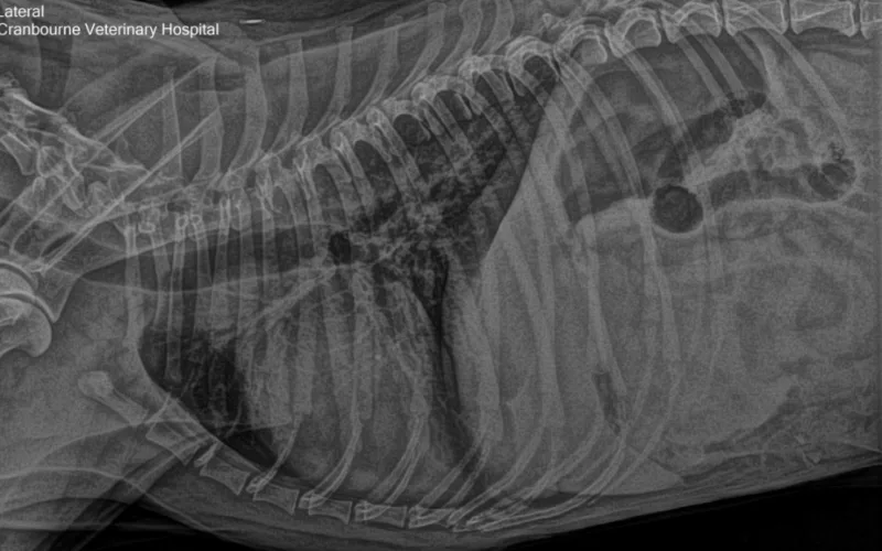
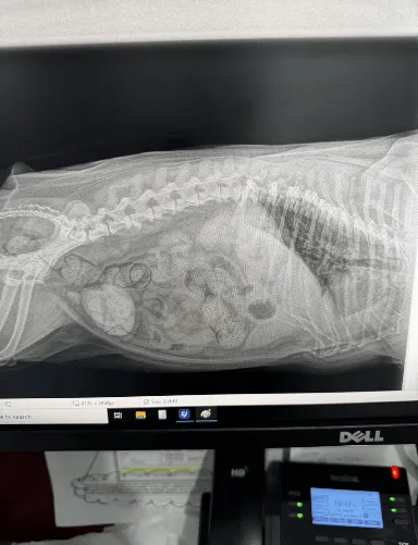
At this point, further imaging was required. Chest X-rays performed at Casey and Cranbourne Veterinary Hospital showed no obvious evidence of tumour spread to the lungs, but the nature of the splenic mass and vascular abnormality remained unclear.
“Diagnosis is not always a single event. New information can emerge as a patient’s condition evolves.”
Bringing Advanced Diagnostics To The Hospital
Given the complexity of the findings, Barney’s case became a collaborative effort involving our veterinary team and external diagnostic imaging providers.
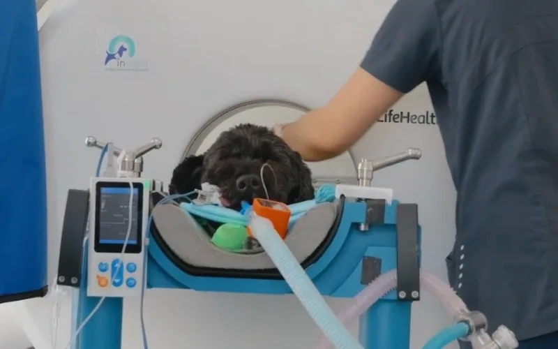
An emergency referral was initially discussed. However, after considering all options, Barney’s family elected to continue monitoring him while we arranged a mobile CT service to attend the hospital. Before proceeding, additional testing was performed to ensure the vascular abnormality was not affecting liver function and would not increase anaesthetic risk.
A bile acid tolerance test confirmed normal liver function, while coagulation testing showed no increased bleeding risk. Several days later, Insight Mobile Veterinary Diagnostics attended Casey and Cranbourne Veterinary Hospital to perform a CT scan.
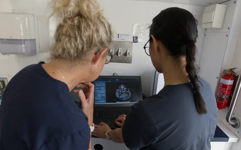
The CT provided the answers we needed. It confirmed the presence of the splenic mass and revealed that the unusual blood vessel connection was a rare congenital variation between the caudal vena cava and the azygous vein. Importantly, this vascular anomaly was not connected to the liver and was considered an incidental finding that did not prevent surgery from proceeding safely. With this information available, a surgical plan could be developed with greater confidence.
Surgery And Histopathology
Barney was taken to surgery by Dr Leanne Versteege, who performed a splenectomy (removal of the spleen). During surgery, evidence of previous bleeding from the splenic lesion was identified. Adhesions had formed between the spleen and surrounding abdominal tissues, where the lesion had partially ruptured and bled.
The spleen was successfully removed, and all associated blood vessels were carefully ligated without complication. The removed tissue was submitted for histopathology. While splenic masses can sometimes represent cancerous conditions, Barney’s results provided welcome news.
The final diagnosis was complex nodular hyperplasia with an associated haematoma. There was no evidence of cancer. With the spleen removed, the risk of future bleeding from the lesion was eliminated.
Recovery And Outcome
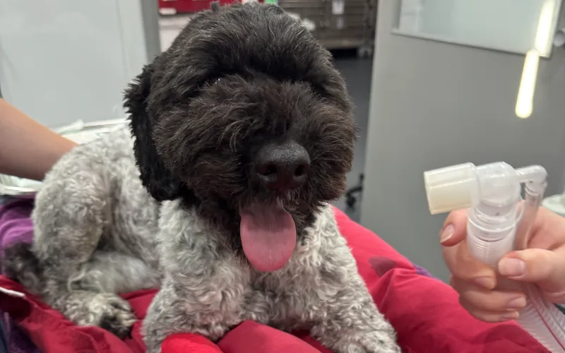
Barney recovered exceptionally well following surgery. In fact, his biggest challenge became staying quiet during the recovery period. Despite requiring strict rest, he quickly returned to his energetic personality and required medication to help keep him calm while healing.
In the weeks following surgery, his family noticed something interesting. Looking back, they felt Barney had probably been quieter and less energetic for several months before becoming acutely unwell. Following the splenectomy, he appeared brighter, more active and much more like himself.
Today, Barney continues to do well and has returned to normal life with his family.
“From the very beginning the care we received was exceptional. The team understood both Barney’s anxieties and our own, supporting us through every step of a very complex situation. All of the vets who helped look after Barney whilst he was very ill were amazing. Leanne, the owner of Casey and Cranbourne Veterinary Hospital, personally performed Barney’s surgery and went above and beyond throughout the entire process. Every single staff member, vets, nurses and admin genuinely cared for Barney, and that compassion made all the difference. We felt completely safe in their hands. We wouldn’t go anywhere else.” – Natalie | Barney’s Owner
Why Barney's Case Was Unique
Barney’s case highlights how multiple conditions can exist simultaneously. He initially presented with signs consistent with pancreatitis, but ongoing assessment and advanced diagnostics uncovered a partially bleeding splenic lesion as well as a rare congenital vascular anomaly. Had these findings not been identified, the splenic haematoma may have progressed to a more significant abdominal bleed.
Cases like Barney’s demonstrate the importance of thorough investigation, collaboration between veterinary professionals, and having access to advanced diagnostic pathways when answers are not immediately obvious.
As Dr Leanne Versteege reflects, one of the most important lessons for pet owners is simple: If your pet is off their food, seems unusually quiet, is vomiting repeatedly, or simply does not seem themselves, trust your instincts and arrange a veterinary assessment. Subtle changes in behaviour can sometimes be the first sign that something more significant is happening.
Our Services
Trusted Veterinary Services All Under One Roof
Your pet’s care is supported by a highly experienced team who provide everything from routine check ups and preventative care, to advanced diagnostics, surgery and dentistry. We take a careful, thorough approach to every case and keep you informed at each step, so your pet receives reliable, compassionate care throughout their life.
Whether your pet needs everyday health support, or help with a more complex condition, our team work together to provide clear answers and thoughtful treatment in a calm, modern hospital.


PET EDUCATION AND RESOURCES
Practical advice and real case stories to support your pet’s care.
Pet Care Articles
Helpful tips and expert guidance to support your pet’s health and everyday well-being.
Real Life Case Studies
Real stories from our veterinary team showing how we diagnose, treat and support pets through illness and recovery.

Quick Answers to Common Questions
Can pancreatitis hide another underlying condition in dogs?
Yes. While pancreatitis can cause vomiting, lethargy and reduced appetite, those symptoms are not unique to the condition. In Barney’s case, ongoing abdominal pain and changes in his blood work prompted further investigation, which ultimately revealed a splenic mass and a rare vascular abnormality.
Why would a vet recommend an ultrasound as well as blood tests?
Blood tests provide valuable information about organ function and inflammation, but they cannot always identify structural abnormalities. Barney’s ultrasound revealed an eight-centimetre splenic mass and abdominal bleeding that could not be detected through blood testing alone.
What causes a splenic mass in dogs?
Splenic masses can develop for several reasons, including benign changes, blood-filled lesions, or cancerous growths. Histopathology is often required to determine the exact diagnosis, which in Barney’s case confirmed complex nodular hyperplasia with a haematoma rather than cancer.
Can a dog live a normal life without a spleen?
Many dogs recover well after a splenectomy and go on to live normal, active lives. Barney returned to his usual energetic personality after surgery and continued to do well during follow-up monitoring.
What is a congenital caudal vena cava anomaly in dogs?
A congenital caudal vena cava anomaly is an uncommon variation in the way major blood vessels develop before birth. Barney’s CT scan showed a connection between the caudal vena cava and the azygous vein, which was determined to be an incidental finding that did not prevent surgery from proceeding safely.
When should vomiting and loss of appetite be treated as an emergency?
Repeated vomiting, refusal to eat for several days, lethargy, abdominal discomfort, or significant behavioural changes should always be assessed promptly. While some cases may involve a relatively straightforward gastrointestinal issue, others can reveal more serious underlying conditions that require urgent investigation.
Book your pet’s appointment
today with our experienced, caring team.

Newsletter sign up
Contact Info
Opening Hours
Monday to Friday: 8:00am –
7:00pm
Saturday: 8:00am – 3:00pm
Sunday: CLOSED
Closed on public holidays
For all out-of-hours emergencies,
please contact Veterinary Referral Hospital (VRH) on 1300 385 874
or Casey Pet Emergency on (03) 8790 1625.